


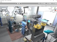

In my project my intention is to create a 3D virtual human anatomy that a medical student can use to earn dissection. If they make a mistake, press a virtual button and a different body appears, so that's why they could dissect again. You could do many times as you want. No harm to anybody, it is a computer model.
Virtual surgery is predominantly going to be used by surgeons in the near future. If it is complicated surgery, even an inexperienced surgeon might want to do a preplanning or rehearsal. This technology could be very useful in that situation. The same tool is also useful when you are training a surgeon. The cycle of training is quite long. By using this kind of techniques, we could have an expert to perform a particular surgery and record all the movements that the expert practised.
We could ask a novice to perform the same operation, and compare the two signatures. Then based on that we could actually hold training plan to say how quickly we could take the novice to the same level as an expert.
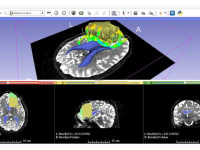
Another focus of this project is to create and develop virtual dissection table. The table is going to be about the size of hospital bed with the top as a touch screen computer. This device is going to be produced from carbon-fiber material, so it's capable of working coordination with X-Rays. Thus we will be able to work on medical scans such as PET,CT, and MRs. So that's why one table could give countless medical students the ability to work with a virtual cadaver, enlarge or rotate biological systems and bisect or remove body parts. Furthermore, another concept that the project deal with is to take a stack of 2D medical scans and extrapolate a 3D model from them. Then the model could be printed out from a 3D - printer. A medical doctor could scan a patient's cancer and then explain with a physical model (with the tumar opaque) exactly what the problem is. Before we print out the 3D model , we make a slight modification : We extrude a series of small slots from the model within the tumor. Then we printed out and put tiny radiography chips into the model. This means that now we have a patient - specific device(model) that we can shoot radiotheraphy at. Medical professionals could use this model to focus the beam and find out more about peripheral damage. The end goal of this process is to improve cancer treatment by developing new techniques on radiotherapy.
On the other hand, some medical school and hospital training programs are going to be simulated by visual reality environment of this application such as some CME (continuing medical education) courses. Thus medical students or inexperienced surgeons are going to be able to develop their medical and clinical skills without doing any harm to any patient due to that virtual enviroment.
Voting
-
ABOUT THE ENTRANT
- Name:Engin Bozkurt
- Type of entry:individual
- Profession:
- Software used for this entry:InVesalius, 3Dslicer, Unity
- Patent status:none