


This invention introduces Time-resolved Hyperspectral Imaging Spectroscopy (THIS), a novel spectroscopic technique for the life sciences.
It can find potential applications in diverse fields, including:
- biohazard monitoring
- rapid mass-screening for infectious diseases
- live-view tumour detection during surgery
- laboratory-based research and pharmaceutical development
- monitoring plant health and parasites in agriculture
Application examples are shown in UK Patent no. GB2609862 (and further pending patent cases).
The invention improves the widely-used technique of Fluorescence-Lifetime Imaging Microscopy (FLIM) by going from the three data dimensions of image (2 dimensions) and fluorescence-decay time (one more dimension) to four, including also the fluorescence-wavelength spectrum in hyperspectral resolution of 100 wavelength channels, or more.
Thus, at each pixel of an image of a live or prepared sample, a fully 2-dimensional data set is provided in the form of hyperspectrally resolved fluorescence spectra and their time dependence. Using readily available technology, the spectral/temporal resolution of the technique can approach the fundamental quantum-uncertainty limit of time vs. photon energy.
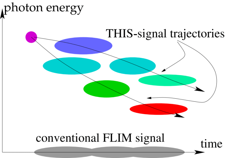
The fluorescence data are then present for each pixel in the form of trajectories in time-wavelength space. This vastly improves the power to discriminate different protein species in a real-world sample because the trajectories in two dimensions are topologically distinct where overlaid fluorescence-decay curves on the time axis from conventional FLIM are not in Figure 1: fluorescence over time and photon energy (reciprocal to wavelength).
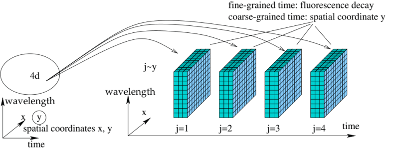
This capability becomes possible with a new generation of high-performance photon detectors such as the LAPPD [1], in combination with a data-multiplexing scheme that physically maps the original four data dimensions onto the three that are natively supported by the detector (in the case of the LAPPD spatially equivalent to about 200 x 200 pixels and timing of each photon to 50 pico-seconds).
The patent describes multiple ways of such multiplexing, but all are based on the realization that fluorescence decay from proteins occurs within nanoseconds, so that the time axis can serve a multiplexed purpose of representing the fluorescence decay with 50-ps resolution over time intervals of 10 to 100 nanoseconds, while successive 10..100 ns time intervals represent another, suitably discretized data dimension. (Figure 2).
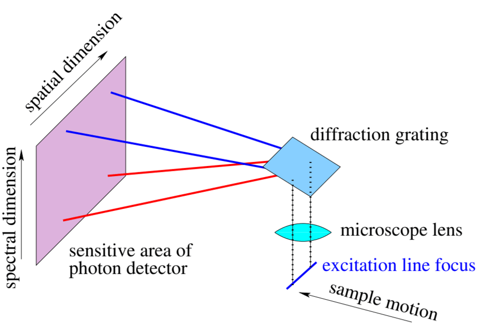
One specific embodiment described in the patent (Fig. 3) is for mass screening in public health. Sample material is pulled through the field of view of a microscope where it is excited to fluorescence by a line focus of a picosecond-pulsed laser. The sample is imaged via a wavelength-dispersive element (grating, prism) onto an LAPPD or other detector capable of continuously reporting photons with 50-s timing and image location.
Then, pixel locations along the line focus correspond directly to a spatial sample coordinate; pixel locations perpendicular to it correspond to wavelength spectrum (in about 200 channels); fine-grained (50 ps) time is the fluorescence decay time, and coarse-grained time (100 ns) is the second sample coordinate due to the sample motion.
Like this entry?
-
About the Entrant
- Name:Bernhard Adams
- Type of entry:individual