


Hydrocephalus, a neurosurgical disorder characterized by an abnormal accumulation of cerebrospinal fluid (CSF) within the brain's ventricles, often requires timely surgical intervention through procedures such as Endoscopic Third Ventriculostomy (ETV) and Ventricular Shunt Placement. These neurosurgical techniques demand a high level of precision and skill, given the intricacy of intracranial anatomy and the proximity of vital structures. To address the critical need for safe, repeatable, and anatomically accurate surgical training, we have developed a Physical Simulator for ETV and Ventricular Shunt Placement, a patented innovation designed to enhance surgical competency outside the clinical environment.
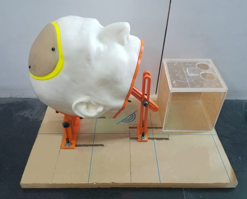
The proposed simulator comprises a modular 3D Printed, anatomically realistic training device featuring a cylindrical housing placed within a fixed anatomical head model. This housing accommodates removable and replaceable artificial anatomical structures that mimic the human scalp, skull, brain, and ventricular system. These components include a soft silicone-based skin patch for incision and suturing practice, a hard bone matrix-based skull patch for drilling and burr hole creation, and a brain and ventricles model made of varied grades of agilus material and silicon-based membrane at the floor of third ventricle to replicate the texture and consistency of actual neural tissues.
Along with ETV, the simulator also offers a dedicated provision for ventricular shunt placement training. The design allows users to create a burr hole, navigate the ventricular system, and insert a catheter for simulated shunt positioning, enabling step-by-step rehearsal of both emergency and elective hydrocephalus management procedures on the same platform.
A key functional highlight is the CSF flow simulation system integrated via inlet and outlet ports on the fixed scalp model, driven by an external motorized pump. This enables realistic visualization of CSF circulation during the procedure, particularly during the endoscopic perforation of the third ventricle’s floor, which is the essential therapeutic step in ETV. Additionally, the simulator provides an anatomically accurate path for inserting an endoscope through a burr hole, navigating intracranial spaces, and executing the critical fenestration task while observing dynamic CSF movement.
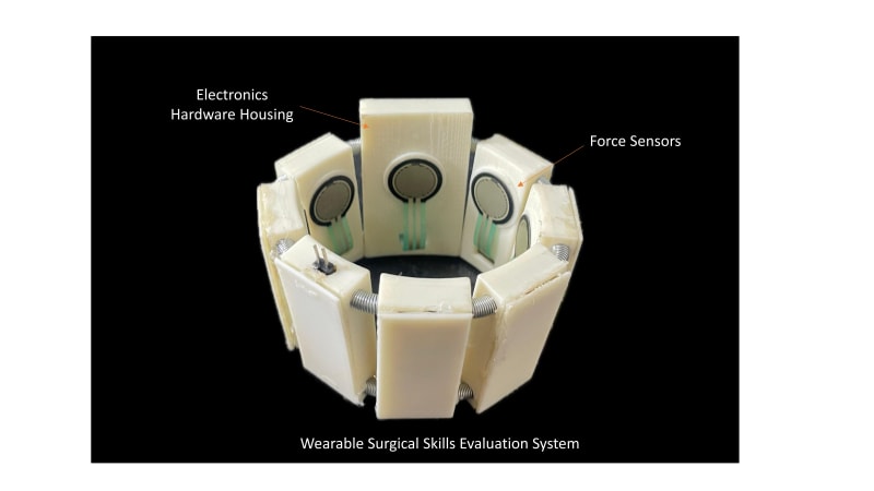
To advance objective surgical skill assessment, the simulator also incorporates a sensorized wearable band embedded with force sensors and inertial measurement unit (IMU) worn by the trainee during simulation exercises. This system records force application patterns, hand movements, and motion dynamics in real time. The collected data is processed using AI-based algorithms to evaluate key performance indicators such as precision, tremor control, task completion time, and procedural smoothness. This feature offers immediate, quantitative feedback to learners and educators, facilitating data-driven skill enhancement and performance benchmarking against expert profiles.
The simulator has undergone validation by experienced neurosurgeons and residents to establish its face and construct validity. It was consistently recognized as a valuable, anatomically accurate, and functionally relevant platform for acquiring and refining neuroendoscopic skills.
Like this entry?
-
About the Entrant
- Name:Dr Ramandeep Singh
- Type of entry:individual
- Software used for this entry:Simpleware ScanIP, NX Unigraphics, Meshmixer