
ADD ON CAMERA FOR TRANESOPHEGEAL ECHOCARDIOLOGY
FIELD OF THE INVENTION
The present invention relates to a medical device for minimally invasive procedures and particularly to an image sensor to be used in such a procedure.
SUMMARY OF THE INVENTION
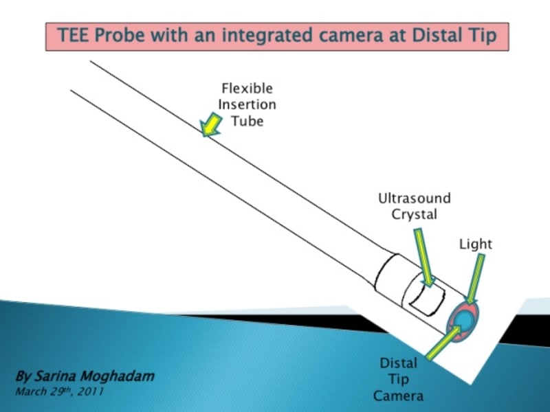
An aspect of some embodiments of the present invention relates to a compact distal tip for mounting on a TEE probe.
This new integrated medical device would reduce risks involve with potential injuries throughout and subsequent to examination of cardiac patient during ultrasound imaging scan and reduce the consequence involve with employ of TEE probe.
BRIEF DESCRIPTION OF THE DRAWINGS
Exemplary non-limiting embodiments of the invention will be described with reference to the following description of the embodiments, in conjunction with the figure. Identical structures, elements or parts which appear in more than one figure are perfectly labeled with the same or similar description, and in which:
BREIF DESCRIPTION OF THE PROBLEM
Since there is no direct visualization of esophagus during TEE (Tranesophageal Echocardiography) probe insertion and manipulations, it requires more attention compared to conventional optical Gastroscope.
Thus conventional TEE studies require increased diligence because of the lack of optical control during probe insertion and manipulation in the esophagus throughout operative procedure.
Example: Often the intraoperative cardiovascular is postponed because of concern for increased esophageal bleeding after heparinization due to linear ulcer adjacent to a stricture of distal linear erosion.
Introduction to TEE:
In comparison with other diagnostic modalities, TEE is relative safe and noninvasive. However, the insertion and manipulation of the ultrasound probe can cause oropharyngeal, esophageal, or gastric trauma.
Implication, potential risks and difficulties:
One proposed source of pharyngeal and esophageal injury during TEE involves improper probe placement. If the tip of the probe is not centered in the posterior pharynx and instead is placed laterally into the pyriform fossa, the probe may bend or “buckle”. Advancement of the probe in this situation may cause the tip to be oriented in an extreme retrograde position. Manipulation or rapid removal of the probe oriented in this manner could cause serious gastroesophageal laceration.
Additional risks and difficulties include:
• False passage into diverticulum
• Inadvertent tracheal intubation
• Probe of TEE can easily slip into Zenkers diverticulum and can cause perforation.
• Forty percent of those perforations occur in the hypopharynx, whereas 20% occurs in the esophagus.
• TEE probe placement, motion and removal may lead to displacement or accidental extubation of endotracheal tube particularly in children.
• Patients with hiatal hernia encounter resistance during probe advancement from the esophagus to the stomach.
• Due to complications associated with conventional TEE probe, often complication is followed by immediate diagnostic upper gastrointestinal endoscopy.
Conclusion:
The true frequency of Esophageal and other related injury associated with conventional TEE probe is undoubtedly underestimated.
A proposed TEE Videoscope is a valuable clinical tool for ease of passage under direct visualization through the hypopharynx into Esophagus, thus it improves patient safety and reduces injury related incidents.
Like this entry?
-
About the Entrant
- Name:Sarina Moghadam
- Type of entry:individual
- Patent status:none