


Non-invasive Imaging Method
A: Electrical Resistance Tomography (ERT) is a non-invasive imaging method applied in industry, geoelectrics during the last decades and recently for medical monitoring and analysis of the human lung functions.
B: What does "non-invasive“ mean?
A: One is able to extract information about the interior of an conducting object (for example the human chest) without measuring on the inside but only on the surface.
B: Similar to Ultrasonic Tomography (UST) and the Computed Tomography (CT)?
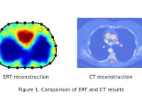
A: Exactly! While UST uses sonic waves and CT uses gamma rays, in ERT the non-invasive character is implemented by means of weak electrical current sources and the measurements of the stimulated electrical potential on the object's surface. Based on that measurement and the mathematical model for the electrical potential stimulation the electrical resistance distribution can be computed for the interior of the given object, see Fig. 1, left.
Low Resolution Problem
A: ERT is a very low-cost imaging method (equipment: ca. 5.000$) as compared to UST (equipment: ca. 15.000-120.000$) and CT (equipment: ca. 100.000-200.000$). At the same time it is absolutely safe to use and provides a very high temporal resolution - up to 1000 fps. But there is one drawback - the spacial resolution ist quite low, see Fig. 1, left.
B: I see, so if the spacial resolution of ERT could be increased, it would be the ideal solution! What are the reasons for the low resolution?
A: Because of the radial spreading of the electrical current in the object one can compare the potential measurement with sampling of small structures using a big brush. That means the information about the small structures in the interior of the examined object gets smoothed pretty much. This smoothing of the data is the main reason for the low spacial resolution.
B: So, can we reduce this annoying smoothing? If so, how?
Solution
A: Using the comparison with the brush, the CT samples the tissue density distribution via a fine, small brush (that is the gamma beam), which is only 0.5mm slim! This of course, leads to a high spacial resolution, as in Fig. 1, right.
B: I suppose your idea is to force the electrones to a beam-like path.
A: Indeed!
B: But how is this possible?!
A: If we extend every electrode by a conducting cap including separated stimulating and meassure electrodes, the domain observed by the measure electrodes reduces to a slim band, see the red band in Fig. 2. In this way the cap forces the electrones into a desired path. Thus, I expect more detailed information about the resistance distribution in the interior of an object.
B: Can ERT compete with UST or even with CT?
A: A spatial chest resolution of 0.5mm as with CT is probably beyond technical possibilities. Nevertheless, ERT could become a challenger for the conventional methods, because a resolution duplication or even triplication would enhance ERT incredible, see Fig. 3.
Like this entry?
-
About the Entrant
- Name:Andreas Schkarbanenko
- Type of entry:individual
- Software used for this entry:COMSOL, MATLAB
- Patent status:none