


Transplantable unions of living cells and electronic circuits interfaced with body parts, will be used to test, measure, monitor, control, repair, replace, or assist the functioality of body parts.
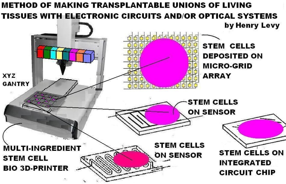
Ordinary cells and stem cells are deposited upon, grown, attached to, united, and coupled with non-rejectable electronic circuits, sensors, radio tranceivers, optical, electro-optical, and/or computer systems, to form new transplantable unions. Cells are 3D-printed, deposited, dipensed, painted, sprayed, air-brushed, injected, squirted, spritzed, or delivered by other means upon circuits, or optical systems.
Circuits can be manufactred by ordinary methods, 3D-printing, or other means. Printed circuits, ICs, other circuits, and optical systems are ordinary or are designed to adaptively couple with cells by having surfaces, undercuts, ridges, depressions, interstitial spaces, bumps, holes, hooks, adhesives, conductive scaffolds, and geometric shapes to which living cells can adhere, or get caught on, within, underneath, and inbetween physically and frictionally.
These unions make thousands of electrical contacts to cellular tissues. These tissues, as well as, wire attached circuits and/or optical systems upon which tissues are grown and coupled with, form unions which are then transplanted into the human body or organs temporarily outside the body.
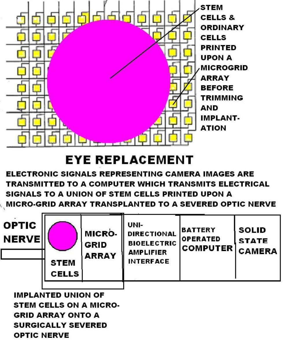
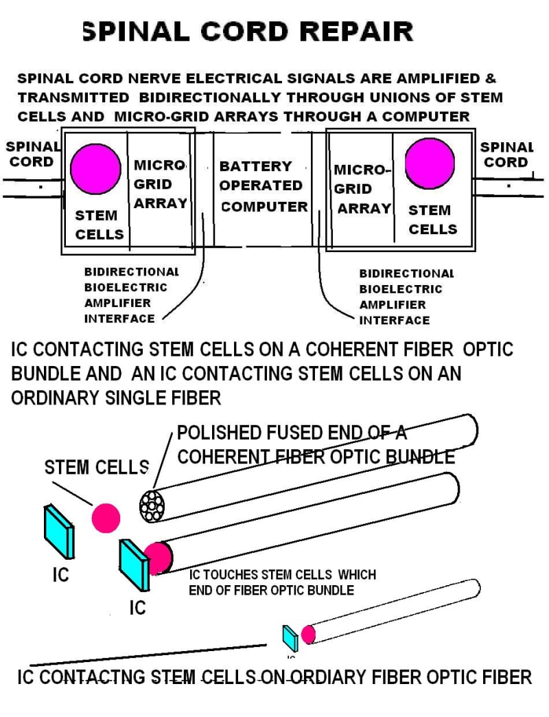
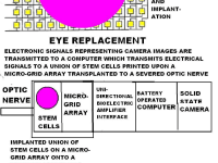
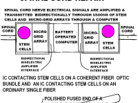
Using this method, the optic nerve, retina, spinal-cord, brain, heart, lungs, liver, kidneys, pancreas, thyroid, blood vessels, muscles, skin, and other human body organs or parts can be connected to, interfaced with , and combined with electromic circuits and optical systems.
Deposition, dispensing, painting, spraying, and filling machines are decades old and are ubiquitous worldwide. Bioprinting cells is well known and widely practiced. Bioprimtimg progressed very fast and greatly since I wrote , “LIVING TISSUE RAPID PROTOTYPING SYSTEM” in the Emhart NASA Create the Future Design Contest in 2006 (Merit Prize Winner), and since I wrote “MULTI RESERVOIR AIRBRUSH LIVING TISSUE 3D STEM CELL PAINTER / INJECTOR,” also published in the NASA Create the Future Design Contest in 2014. Today, ordinary cells and stem cells are cultivated, separated, and 3D-printed by bioprinters on flat or shaped surfaces, structural scaffolds derived from animals, or 3D-printed scaffolds.
Implanted electrodes measure or control bodily functions but have problems. Printed circuits and ICs have been manufactured for more than 50 years and are positioned to touch the skin or implanted within the human body internal organs, and other body parts.
Electrcal surface contact effectiveness for transmitting, receiving, and measuring signals isn’t as good as electrode implantation, but electrode implantation is labor intensive, destructive, and produces scar-tissue (even with micro-fine wire electrodes). Scar-tissue is the body’s method of self-repair. Circuits and electrodes inserted within cut cellukar tissues, severed organs, or body parts, trigger scar-tissue formation there which interferes with bodily function, electrical conductivity, electrical transmitivity, nerve signal transmission, measurement, and control.
Electrodes of electroencephalographs, polygraphs, and eletrocardiographs are attached to skin via gels and adhesives, but they don’t touch many points per unit surface area.
Transplantable unions of selected specialized stem cells on circuits differentiate and grow ttogether with adjacent cells of the body wherever they are transplanted to solve the above described historical problems.
Like this entry?
-
About the Entrant
- Name:Henry Levy
- Type of entry:individual
- Patent status:pending