


How can a medical professional deliver better patient care and increase positive functional outcomes for orthopedic patients? Through better understanding of the underlying biomechanical issue(s). If a physician has a greater understanding of the particular dysfunction, not only can they develop more effective treatment plans, they can convey the issue and treatment options more effectively to the patient. The human body is an elegant machine, we take for granted the complexities inherent in every motion we under take. Through a blending of mathematics and 3D imagery, complex normal and abnormal biomechanics can be visualized for medical professional training,communication and understanding of injury and conveyance of patient treatment options.
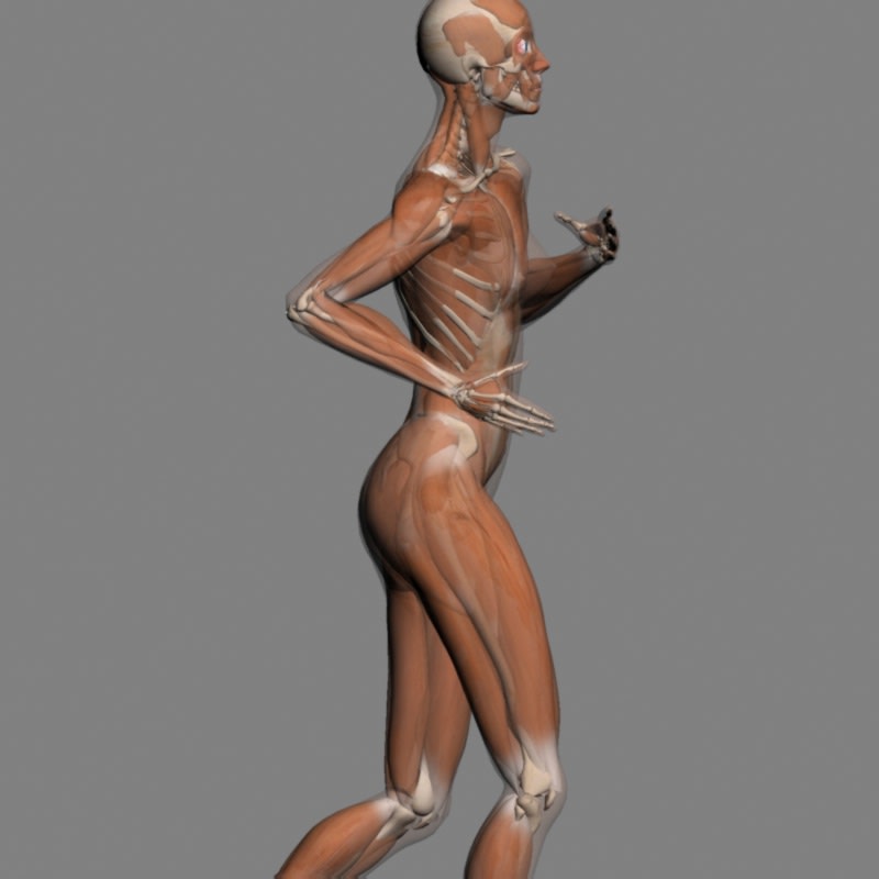
Utilizing the toolset and applied biomechanical algorithms, every muscle, bone and soft tissue structure can be animated to convey biomechanical motion within the fully articulated 3D system. For example, in heel pain syndrome the root structures that are painful and under load are the intrinsic musculature. The subtalar joint pronates and the midtarsal joint axes converge to a more parallel position. This produces an instability within the foot posture leading to greater strain on the biomechanical system and contributes to injury. A visual representation of this issue delivers greater understanding of the contributing factors leading to injury and the development of specific treatment programs that address the underlying issues.
Medical professionals learn from static anatomical tools. When evaluating a patient they are extrapolating this knowledge to a fully articulated moving object with 3 planes of motion. Understanding normal and abnormal biomechanics is difficult without using an animated 3D biomechanics system. This toolset allows the creation of imagery and video representation of these anatomical systems and can be delivered via portable computing devices such as smart phones and the Ipad. This allows the medical professional to convey the mechanism and structure of injury to the patient. Better information regarding the medical issue will lead to the development of more effective treatment plans. Greater patient understanding of their particular medical issue will result in greater compliance to treatment programs and educated decisions regarding their treatment options.
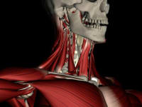
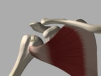
Based on volumetric body data (patented) and a body volume visualization (patent pending), an understanding of muscle dynamics and muscle insertion/origins, a complete biomechanical system is applied to represent a specific medical condition. The end result is a visual representation of the specific issue. The resulting imagery is reviewed by a physician expert, in the associated clinical area, for accuracy. This information is then delivered on demand through a global content delivery system, designed by the inventor of the technology, to the physician attending to a patient or the medical professional training in a particular clinical area. Licensed medical professionals across the world can access this information 24/7 with explanation in their native language.
Better medical information helps deliver better patient care.
See the system in motion
http://www.youtube.com/watch?v=WTERYaLu2QQ
Like this entry?
-
About the Entrant
- Name:Tom Vastano
- Type of entry:individual
- Software used for this entry:3dsmax, Spacepilot, motionbuilder, mudbox
- Patent status:none