
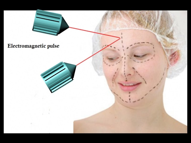
Mitotic Spindles (microtubules attached to the mitotic spindle and to part of the centromere) determines position of divided cells. two structures called centrosomes move to opposite sides of the cell during this phase and begin building the mitotic spindle. String like structures called microtubules grow out from the spindle and connect to the sister chromatids at their kinetochores; one microtubule from one side of the spindle attaches to one sister chromatid in each chromosome, and one microtubule from the other side of the spindle attaches to the other sister chromatid. cell may contain a pair of centrioles (or microtubule organizing centers in plants) both of which are organizational sites for microtubules. Centrioles begin moving to opposite ends of the cell and fibers extend from the centromeres. Some fibers cross the cell to form the mitotic spindle. paired chromosomes separate at the kinetochores and move to opposite sides of the cell. Motion results from a combination of kinetochore movement along the spindle microtubules and through the physical interaction of polar microtubules. if spindle is positioned there by electrochemical position we can alter positions of individual spindles by external electromagnetic force and make new shape for cell structure like human body parts. For a growing child we study growing mechanism to adulthood and make alteration at the right position at right time to get desired shape at adult hood. For an adult human we can stimulate cell division and reposition spindles by electromagnetic force to get desired shape. For reshaping bones in adult we can use the mechanism of bone fracture healing to reshape bones in adult. For that we can use electron scanning by multiple beams and make change spindles position by a small amount by first beam so that we do not disturb neighboring cells much and then progressively by next beam and finalize shape by final beam.
We can use this system to cure internal injury also by accelerating healing process or clot blood. By replacing the coding gene that determines position of spindle with new gene we can make this new shape hereditary. We can use electromagnetic wave to manipulate the position of these spindles. We can use nano electromagnetic or nano capacitive devices to relocate these spindles by magnetic or electric field respectively. to study cell division current flow, place cell on a capacitive surface similar to capacitive touch screen and acquire signals from that to a computer display. You can place capacitive surface on all four sides of human to get 3D image of electric current flow. we should use extremely high sensitivity and super fine resolution capacitive surface. Principle of this concept is electric charge of human body cells and electric charge of capacitive plate produces a capacitance by measuring this capacitance we can identify location of electric charges in human body just like capacitive touch screen. Using this we can study nervous system functioning and other body functions like heart beats.
Like this entry?
-
About the Entrant
- Name:Diji Jayakaran
- Type of entry:individual
- Patent status:pending