


Longer life expectancy in conjunction with improved knowledge of cancers has resulted in increased demands for core needle biopsies. Additionally, cancer diagnosis typically occurs earlier and hence biopsy specimens are required from smaller tissue masses.
Image guidance using ultrasound imaging is frequently used for locating the increased number of smaller suspected tumours. However, ultrasound images do not have the clarity of other medical images (such as X-ray) and the visibility of the needles in such images is generally very poor - they appear like grey glimmers within a flickering grey background. Additionally, during their insertion, it is common for needles to deflect from their intended track thereby producing further uncertainty of the tip position from where the biopsy specimen is collected. For some needle-based procedures, it can take an experienced Interventional Radiologist more than 30 minutes just to ensure that the needle is in the correct location before treatment is started.
This innovation is targeted at ensuring the visibility and positional accuracy of the biopsy needles.
Approximately 30% of core needle biopsies have to be repeated. This is attributable to various factors, a key one being that the biopsy specimen was not taken from the intended region (due to lack of visibility and needle deflection during insertion).
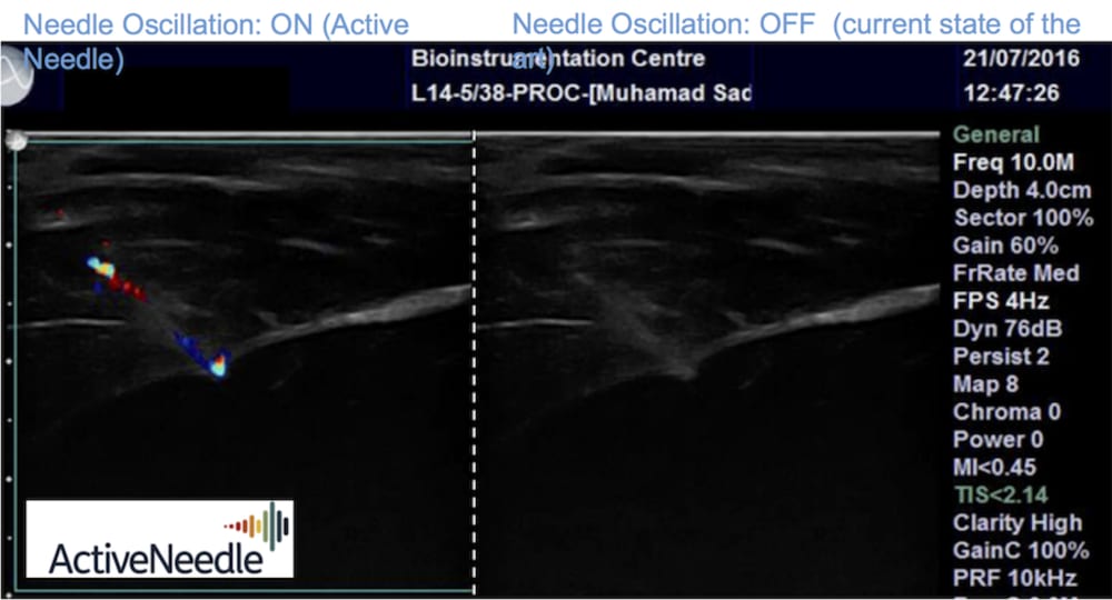
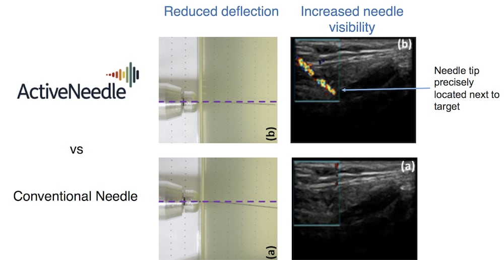
A basic and simple principle of physics has been used to innovatively improve the visibility and thereby de-skill the introduction of such needles. A high-frequency oscillation is applied to the needles resulting in them vibrating backwards and forwards by microscopic distances. The oscillations show up very brightly when imaged in Doppler Ultrasound mode (a mode that exists on ultrasound imaging systems and which detects movement). The coloured Doppler image precisely locates the in-dwelling needle. Optimisation of the way in which the oscillation is applied, reduces the needle deflection. Consequently, this simple innovation provides significant technological advancement by addressing the principal causes of needles being 'incorrectly' located in patients.
Realisation of this innovation is achieved by placing an oscillating transducer in contact with the biopsy needle. The transducer is driven, monitored and controlled by a simple electronic generator. No special manufacturing processes are required and the design is currently being assessed for compliance with international regulatory requirements.
By improving the visibility of the needles and reducing their deflection, we anticipate measurable improvements to key parameters including; duration of procedures, number of repeat procedures, procedure cost, delay to patient's treatment and finally, patient anxiety and well-being.
Halving the rate of repeat biopsies is being targeted. This anticipates savings to healthcare provider of >$300Mn in USA and Europe. This value does not include the reduced cost and improved quality of life for those patients who do not experience the delay to subsequent treatment that is the result of needing a repeat biopsy.
Finally, the technology is readily applicable to other interventional medical procedures. Taking some of these in to account provides a conservative market potential exceeding $1Bn.
-
Awards
-
 2017 Medical Honorable Mention
 -
 2017 Top 100 Entries
Like this entry?
-
About the Entrant
- Name:Mike Irivne
- Type of entry:teamTeam members:Dr Muhammad Sadiq Ian Quirk Dr Mike Irvine
- Software used for this entry:3D CAD, 3D modelling, FEA
- Patent status:pending