
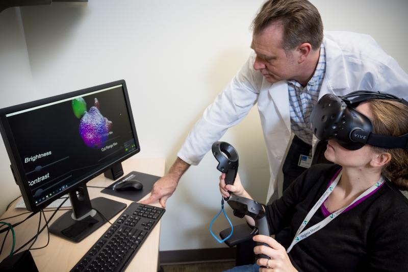
This design uses virtual reality (VR) tools to conduct detailed examinations of human melanoma and disease progression and possible treatment. The tests will use a ConfocalVR systems. Instead of viewing cell images as a three-dimensional model on a flat computer screen, clinicians and researchers will be able to project cell images from normal and diseased tissue into a VR space, see detailed images of internal structure and directly interact with the three-dimensional images in virtual reality.
Patients themselves can immerse themselves in the VR space and view the VR images using our tools. ImageJ software free from the National Institutes of Health (NIH) is used to convert confocal microscope image stacks into 3D volumetric object files (NIfTI ) that can be viewed. Confocal microscope image-stacks are fully immersive in virtual reality compatible with HTC Vive, Oculus RIft, and Microsoft Mixed Reality. The system may also be used by pharmaceutical companies to conduct clinical trials and translate lab discoveries into real-life personal medicine applications using VR tools.
Like this entry?
-
About the Entrant
- Name:Joseph Tarsio
- Type of entry:individual
- Patent status:pending