


Endoscopes have evolved into highly specialized devices that cost tens of thousands of dollars to purchase and to maintain, yet the complexities of these advanced imaging and surgical access devices obviate sterilization between uses; such devices may be disinfected between patients but are damaged by true sterilization while cross-contamination between patients has caused severe trauma and has even led to multiple deaths. As one can imagine, space is a premium in endoscopy, where the working tips of devices like duodenoscopes and ureteroscopes house components for target area illumination (optical fibers or surface mounted LEDs), imaging (video chips or imaging fiber optic bundles), irrigation (open lumen for saline flow) and instrument access (open lumen for passing instruments such as laser energy delivery fibers, forceps, baskets) and steering (control wire anchors) within 2 mm diameters. Compromises are made in providing these capabilities within such small volumes; lighting is typically suboptimal, irrigation flow is slow, image resolution is minimal and the tools that can pass the working channels are limited in capabilities.
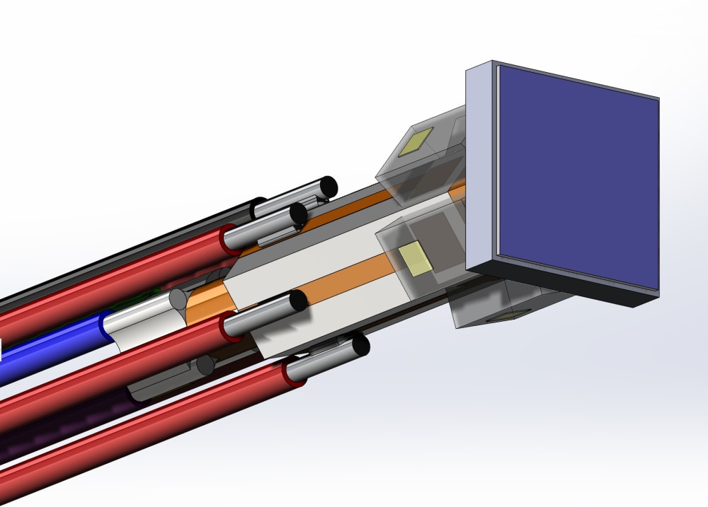
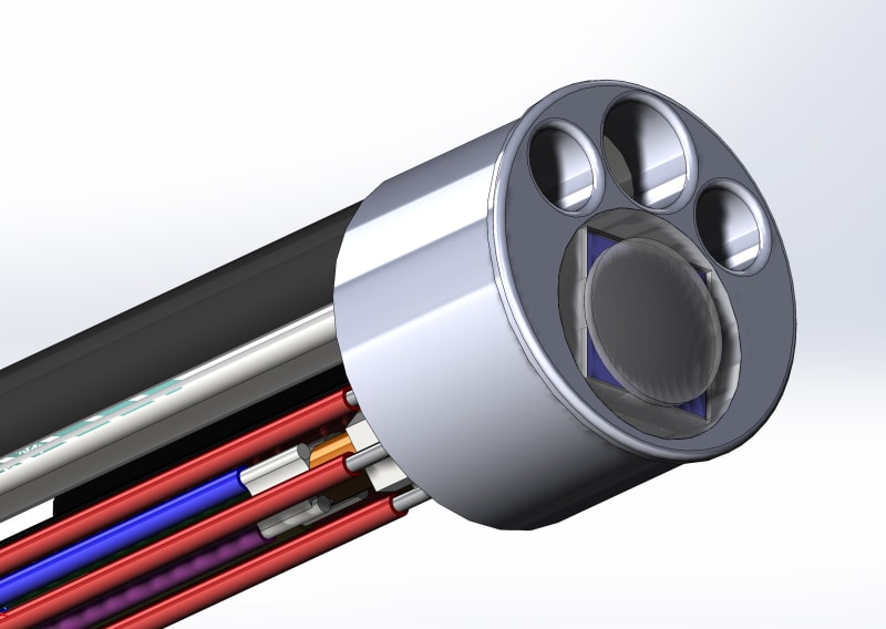
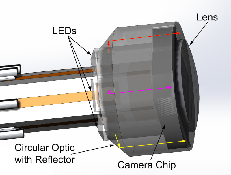
The miniaturization of LEDs, offered in multiple wavelengths, combined with advancements in micro-optics and low-cost grayscale imagers, has permitted the development of illumination schemes for tiny video chips that require less space than color chips, enable smaller profiles and more flexible scope tips within disposable devices. Larger working and irrigation channels may be afforded while multicolor illumination provides a capacity for several fold higher (false color image) resolution than existing devices. The black and white imaging chips and color LEDs are herein arranged such that a dozen or more LEDs may be stacked behind the camera chip, in a space-saving arrangement that also provides for more uniform illumination of the image field via output profile smoothing micro-optics. Small electrical conductors provide both the low voltage power for the camera and LEDs and carry sequential monochromatic image information back to a reusable power supply and image processor located outside of the disposable scope length.
Hyperspectral images may be produced through sequential illumination of the image field with different colors, including wavelengths residing outside of the visual spectrum, enabling imaging of structures like blood vessels and nerve bundles residing below the ‘visible’ tissue surface while enabling further techniques such as light scattering and fluorescence spectroscopy for optical biopsies and monitoring surgical progress and temperature. One can envision the use of two image chips, slightly separated, for producing three-dimensional images.
-
Awards
-
 2022 Top 100 Entries
Like this entry?
-
About the Entrant
- Name:Stephen Griffin
- Type of entry:individual
- Software used for this entry:Solidworks
- Patent status:patented